Object
To search the swelling of DNA hydrogel
Beforehand of making spider-web hydrogel swell, we evaluated swelling behavior. To do this, we use DNA gel beads we generated in Experiment2. This hydrogel beads enable us to analyze hydrogel movement easily.
Protocol
First we prepared DNA gel beads (Tube1) and j2 (Tube2) solutions shown in Table1. We put Tube1 in a well plate respectively and we observed that hydrogels kept their shapes in TNE buffer by microscope imaging. In order to visualize the chemical activity of j2, part of j2 are modified Cy5.
After observing DNA hydrogel in TNE buffer, we put continuously solution of Tube 2 to start to swell the gels. In order to check the final ratio and the speed of swelling, we put different concentrations of j2 for every sample.
We observed DNA hydrogel beads by fluorescence imaging on FAM channel. A part of st1 is modified FAM moieties.
Table1. Materials of experiment 3 swelling DNA hydrogel

Result
In figure 3-1 to 3-6, we show the images of swelling hydrogels (3-1 Channel Cy5, 3-2 to 3-6 Channel FAM).


Left: Figure 3-1. Swelling gel image(Cy5) [j2] = 17 µm, Right: Figure 3-2 ~ 3-6. Swelling gel image(FAM) [j2] = 67, 33, 17, 8.3, 0 µm, respectively.
Right after adding j2 to gel beads, hydrogel started swelling. The growth of sample [j2] = 67, 33, 17 µM stopped around 30 min. On the other hand, control showed almost no expansion.
To analyze the swelling of DNA hydrogel beads quantitively, we measured area of each DNA hydrogel from the movie. Then we calculated relative values of area of each hydrogel bead to the first image for every stack, plotting in Figure 3-7
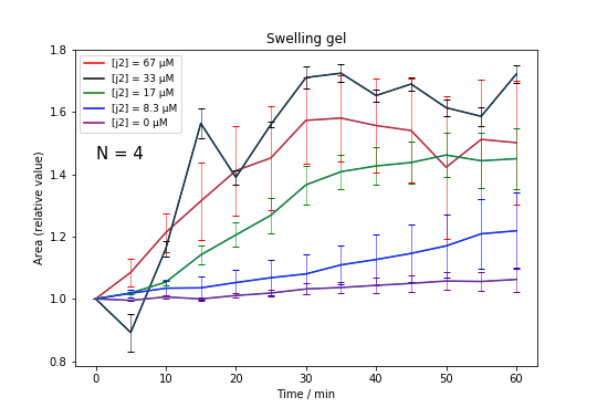
Figure. 3-7 Area(relative value) with time scale, error bar shows standard deviation
According to the samples of [j2] = 67, 33, the maximum value of swelling ratio would be among x1.6~1.8. Additionally, swelling speed depended on the concentration of j2. Sample [j2] = 8.3 µM have continued swelling at 60 min.
Discussion
According to the microscopic movie, we confirmed that the DNA hydrogel beads apparently swelled with adding j2. Cy5 modified j2 attached to DNA hydrogel and make it brighter, this suggests that j2 can hybridize with DNA hydrogel structures. From the result, the speed of swelling seemed to be correlates to concentration of additional [j2].
The maximum size of swelled gel was around 1.6 times because the growth of [j2] = 17 ~ 67 µM was saturated around 1.6 times.
Theoretically, size of a mesh which is constructed by X-motif becomes 1.85 times by extending stem loop structure, and we expected the area of mesh becomes 3.42 times larger. Observed value was smaller than expected.
One of the considerable reasons is that j2 could not enter the core of hydrogel, but just around surface. If we observed longer time scale, the hydrogel would have swelled in higher ratio.