Object
To see if we can shrink the DNA hydrogel after the swelling of the gel
In Experiment 3, we revealed that the shrinking movement of DNA hydrogel occurs with adding j2. Before trying to shrink the spider web hydrogel, we evaluated the shrinking behavior quantitatively by using DNA hydrogel beads. We first put j3 to the DNA hydrogel in the well plate to expand DNA hydrogel. After 1.5 hours we put j3', which peels j3 from l1-j3 hybridization to check the shrinking of the DNA hydrogel.
Protocol
The protocol of swelling and shrinking the gel is shown in Table2.
Table2. Materials of experiment 4 swelling and shrinking the DNA hydrogel
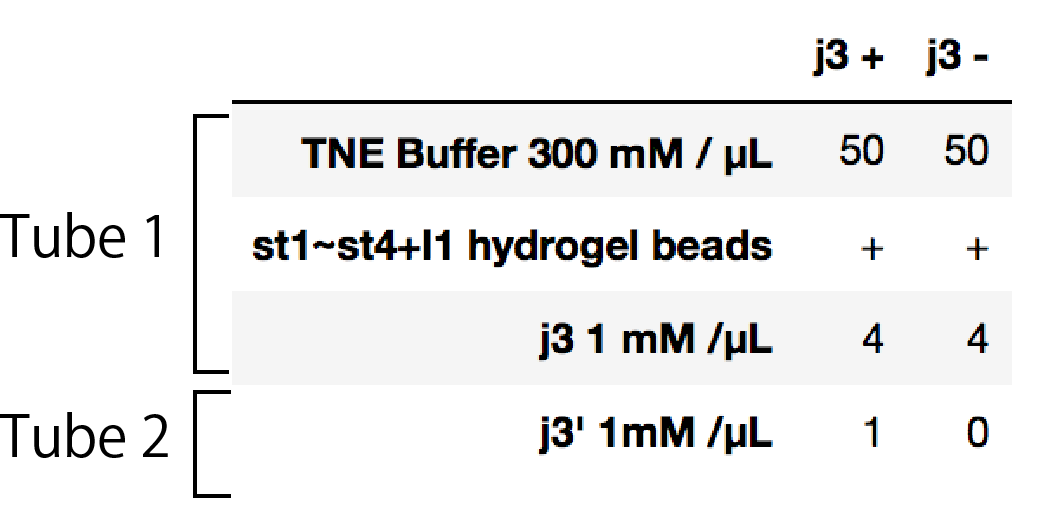
We prepared two tubes for this experiment. Tube1 contains DNA hydrogel, buffer and j3. Tube2 just contains j3'.
First, we prepared tube1 contains in well plate, because gel swelling would start right after adding j3, we start imaging as soon as putting j3. We took the figure of gels every 5mins. After 90mins, we put j3' and continued imaging. this time, we captured every 1minute because of shrinking speed written after.
Result
In figure 4-1 and figure 4-2, the images of the shrinking hydrogels are shown (Channel FAM).
Figure 4-1. Left:Shrinking gel image(FAM) j3+ Right: Control gel image(FAM) j3 -
According to Figure 4-1, we can see that j3 can also make the gel swell like j2 did. After confirming the gel swelling, we put j3'. The movie of the gel shrinking after putting j3' is shown in Figure4-2.

Figure 4-2. Left:Shrinking gel image(FAM) j3+ Right: Control gel image(FAM) j3 -
As we can see in Figure4-2, the gel immediately shrunk after putting j3'. To analyze quantitively, we measured the area of the DNA hydrogel from the movie. Then we calculated relative values of the area of each hydrogel bead to the first image for every stack, plotting in Figure 4-3. Red Shows j3+ hydrogels and black shows control hydrogels.
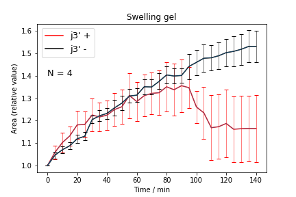
Figure. 4-3 Area(relative value) with time scale, error bar shows standard deviation
With adding j3', hydrogel shrank almost 0.85 times(1.3 → 1.1, relative area) rapidly, although the gels without j3' hydrogel continued to swelling. After j3' added, hydrogel stopped to shrink for 20 min and then kept their areas for end of experiment.
Discussion
The DNA hydrogel apparently shrank 0.85 times with j3'. Shrinking speed was faster than that of swelling. We are succeeded in generation of DNA hydrogel droplets which swell and shrink triggered by single strands, j3 and j3'.
After the gel stoped shrinking, the size of hydrogel was larger than before. We considered two reasons about this result. First, this implies j3' could not removed all j3 from hydrogel. Second, the structure of hydrogel after shrinking might change from before swelling. There can be considered because of room temperature incubation, there might be some non-spesific binding of DNA which cause the sides of gel smaller. With adding j3, the specific binding around l1 is once removed, then adding j3' it would form binding as we can expected.